

Table of contents
Blurred Ultrasound Diagnosis: You know the feeling of intense frustration.
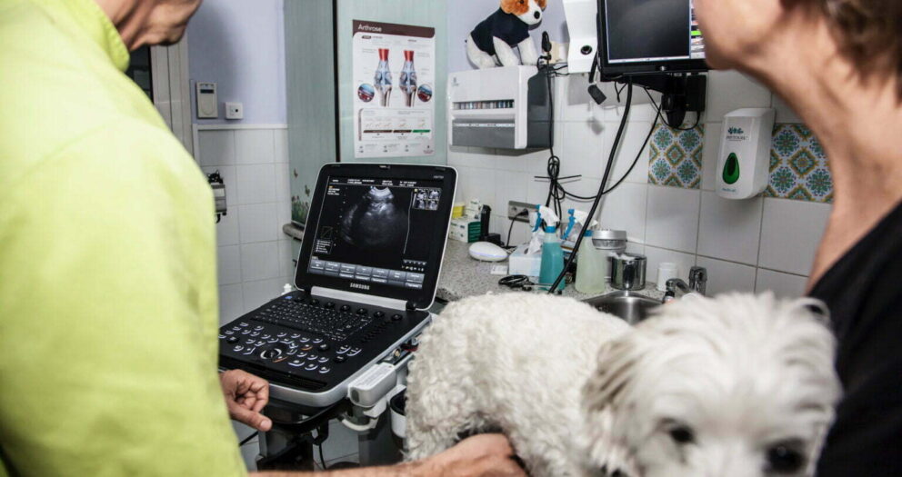
You're in the middle of an abdominal exploration on a dysorexic cat. The room is plunged into darkness, you squint at the screen, play with the gain and focal length, but nothing helps: the image remains gray, grainy, uncertain.
Is it a thickening of the intestinal wall or a simple reverberation artifact? Is it the beginning of a pyometra or a normal uterine horn?
This unclear ultrasound diagnosis is not inevitable. It is often the symptom of a faulty imaging chain. Working “blind” is the royal road to medical error, and the consequences vary according to your location.
1. Blurred Ultrasound Diagnosis: The Double Risk France vs Africa
Faulty equipment is unforgivable, but the penalty varies depending on your continent.
THE GEOGRAPHIC BRIDGE: THE REAL IMPACT
In France (Le Risque Juridique) :
The modern homeowner is a litigious person. If you miss a foreign body because of a mediocre image, you'll be held liable for “loss of chance”. The courts require an obligation of means: using a 2010 ultrasound scanner in 2026 may be considered a technical fault.
In Africa (Reputation Risk) :
Here, the court is WhatsApp. If you send a bitch home with “nothing to report” and she comes back 48 hours later with an emergency pyometer to a colleague equipped with a new Chison ultrasound scanner (see range), your reputation is instantly destroyed throughout the neighborhood.
2. Autopsy of a degraded image: Why is your probe “dead”?
Many vets think that the ultrasound machine is to blame, whereas the culprit is often in their own hands. Probes don't last forever, especially in our climate.
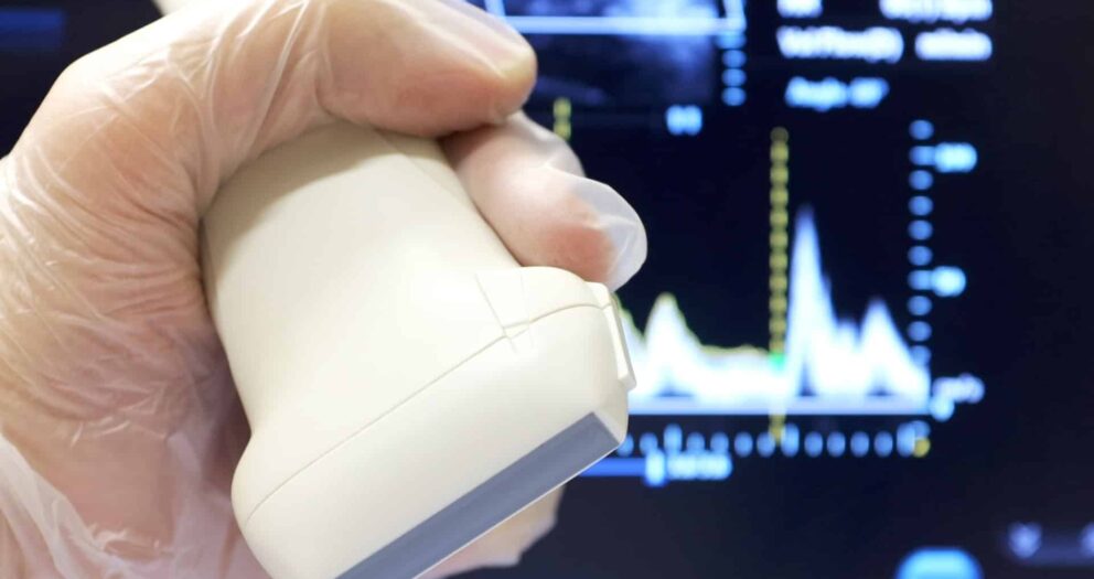
A. Delamination of the Matching Layer
Look at the head of your probe (the grey eraser). Is it swollen? Loose around the edges?
This layer serves to adapt the acoustic impedance. If it becomes detached (often due to tropical heat and humidity), the ultrasound no longer passes through. The result is a unclear ultrasound diagnosis immediate.
If you are still unsure about which type of probe to buy (Convex or Linear?), consult our technical guide : Black & White or Doppler: What's really essential?.
B. The “Death” of piezoelectric crystals
A probe contains hundreds of crystals. With impact, they crack, creating vertical black lines (drop-out lines). The processor tries to “smooth out” these holes, creating a droopy image that masks small details such as linear foreign bodies.
3. The Anatomy of Medical Risk
The danger is not in “seeing nothing”, but in “misinterpreting”.
- Missed Hemoabdomen: A small hypoechoic splenic mass may be invisible on an old screen, drowned out by the “noise”.
- The Pyrometer / Hydrometer distinction : Without clear definition, you risk operating for nothing.
This is where the financial issue often gets in the way of renewal. “Change is expensive”. This is not true. We have calculated that a modern device can be reimbursed with just a few procedures per month.
Check the calculation for yourself here : Veterinary profitability: How to pay off an ultrasound scanner in less than 12 months.
4. The Technical Solution: Going Portable
Technology has taken a giant leap forward. Modern portable ultrasound scanners incorporate DSP processors that clean up the image in real time.
- Speckle reduction: The image is smooth, the liquids anechoic black.
- Tissue harmonics (THI): The contrast between the edges of the organs is accentuated.
Don't let obsolete equipment put your patients at risk.
Free audit of your probes
In doubt about the condition of your crystals?
Send a photo of your image (frozen screen) to an expert Nimedix Medical on WhatsApp.
We'll tell you instantly if the problem is with your settings or if your probe is clinically dead.


Write a comment
You must be logged in to post a comment.